DRAFT WORK PRODUCT SUBJECT TO CHANGE OR DELETION
Alleged date of injury: December 16, 2023
Which days did you see Mr. Atkinson?
October 15, 2024, December 3, 2024, January 17, 2025, with the OCT pages and the “Traumatic Optic Atrophy” assessment.
October 15, 2024 (First visit)
Apparently another ophthalmologist, Forrest Murphy MD, referred Mr. Atkinson to you because he was “unsure of the etiology of decreased vision” in the left eye of Mr. Atkinson and recommended that he see you for a second opinion (Azul vision – May 30, 2024). Is that correct?
Why was Dr. Murphy perplexed?
– Why did you write that Mr. Atkinson had blurred vision “since trauma of glass entering the eye on December 16, 2023?
– Was your notation of trauma based on the word “trauma” that was provided by Mr. Atkinson or was it derived from any record of actual trauma that was described in the medical records?
– Did you review any medical records from Eisenhower immediately following the alleged injury?
– Are you aware that he had an eye examination after the injury that did not show glass in the eye, corneal abrasions, or glass injury to the eye?
– Mr. Atkinson had a history of nuclear cataract in each eye?
– Were his cataracts related to the incident on December 16, 2023?
– What was his uncorrected vision (OD=20/40, OS =CF)
– So he could not even read the eye chart with the left eye? (yes)
– Was his pupil testing normal? Did he have an APD in either eye?
– What is an APD or a RAPD? What is its significance?
– Would you expect to see an APD with an optic nerve injury that causes visual loss to CF? (yes)
– Did you observe the presence of cataract in each eye? (Yes)
– You also noted that the optic nerve had no edema? Why is that significant?
– You noted that there was no disc pallor of the optic nerve in both eyes. Why is that significant?
– Did he have any signs of inflammation in the eye? (No)
– The record states that the OCT was normal. Why is that significant? (the retina and optic nerve are healthy)
– What were the results of a CT scan of the head and orbits? Normal
– Did the scans show any evidence of an injury to either eye? Did they show glass in either eye? (No)
– Why did you write “Unsure etiology of decrease in vision”?
– You wrote rule-out CRAO (central retinal artery occlusion) & cerebral vascular accident? (Yes)
– Why? (concern with a possible stroke). A stroke can happen without glass getting into the eye? (Yes) Can glass on the surface of the eye cause a stroke? (No) What does a work-up for a vascular occlusion or accident consist of (vascular angiography). Was that performed? (No)
– Was any type of treatment recommended? (No)
– So you recommend that he follow-up in 5-6 weeks to have visual field testing. Why? (test to rule-out stroke involving the visual system)
December 3, 2024
This was the second time that Mr. Atkinson saw you? (Yes)
The record shows that you tested his vision, measured his eye pressures, and performed keratometry (Yes). What was the vision in his left eye (CF at his face) Did you examine his optic nerves (no).
The record shows that you requested an MRI head scan with orbits without contrast. You also wrote “Optic Atrophy, OS”. Was the diagnosis of optic atrophy something that you diagnosed without performing an examination of the optic nerve, or was it just a tentative label for ruling out optic nerve damage and justifing exploration with a radiologic study? So you planned to see Mr. Atkinson after an MRI? (Yes)
January 17, 2025
Was this was the third time you saw Mr. Atkinson? (Yes)
What did Mr. Atkinson tell you about his vision? (He said it was decreasing)
What was his vision on this day (CF at 2 feet) and how did it compare with his previous visit (CF at face)? It was improved compared with previous visit).
What did testing of his pupils show? (Normal reactions to light).
Was an APD present in either eye (No).
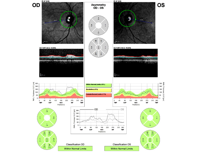
You also performed OCT testing with each eye. What did that show you?
It showed that the average RNFL (retinal nerve fiber layer) thickness was 127 microns for the right eye and 91 microns for the left eye.
Was that significant for progression of optic nerve atrophy in the left eye?
(No. Compare with OCT taken Feb 2, 2024: The RNFL for the right eye was 123 and for the left eye it was 86. Both measurements are “Within Normal Limits”
The same conclusion when compared with OCT readings taken 2/21/2024.
(the OCT images from February 8, 2024 were interpreted by Dr. Forrest Murphy as having no thinning any thickness readings of 100 microns, OD, and 94 microns, OS. The interpretation was described in the record but the actual printout was no reproduced in the record).
Are you interpreting the readings from January 17, 2025 to be consistent with optic atrophy? (No).
What was the condition of the optic nerve of his left eye when you examined him on this visit? (No record of the optic nerve being examined on this visit)
But your diagnosis on this visit was “Traumatic optic atrophy OS secondary to contusion of the optic nerve”.
Is a contusion a soft-tissue injury by blunt force that causes swelling of tissue?
Where in the record is there evidence of blunt trauma to the optic nerve? (No).Was there optic nerve edema or swelling when you first examined Mr. Atkinson about 10 months after the alleged injury? (No. Chart states no disc edema). Did the CT scan show any evidence of swelling of the face or optic nerve? (No)
On this date did Mr. Atkinson have an APD in either eye (No). Is that consistent with a normal optic nerve in each eye? (Yes)
What evidence from the record is responsible for characterizing the alleged injury as a “contusion”?
Would you agree with other ophthalmologists on this case who said that Mr. Atkinson had no identifiable reason that explained his alleged loss of vision in his left eye?
Additional Records Review
The records demonstrate that automated visual-field testing was attempted and repeatedly produced markedly abnormal results, but the studies are too unreliable to establish a reproducible altitudinal defect or to use the field pattern confidently to diagnose anterior ischemic optic neuropathy.
2/13/2026
Dx: Ischemic Optic Neuropathy (Classically not related to trauma)
Main causes: Systemic Hypertension, hyperlipidemia, Diabetes.
3/4/2025: Dr. Ciampa, O.D.
Dx: Disc pallor, OS. But no APD.
“Explained condition does not have a cure
“Doctor, how do you reconcile hand-motion vision from severe unilateral optic atrophy with the absence of an RAPD when the fellow optic nerve was clinically normal?”
“What objective finding established that the optic atrophy resulted from a contusion of the optic nerve, as opposed to another cause of optic atrophy?”