Forensic analysis can provide details regarding the presence of past ocular problems and treatment. This case illustrates how analysis of medical records and adequate documentation may be used to acquire an accurate assessment.
73 year-old-woman gave a history of recent abrupt vision loss following a motor vehicle accident. The woman reported no history of vision problems prior to the accident.
Medical records showed that the patient had a recent history of gradually declining visual acuity in both eyes. The patient has a history of central retinal vein occlusion about one year prior to retinal fluorescein angiography. The patient was taking Lumigan and Betoptic eyedrops to control intraocular pressure.
Ocular exam shows visual acuities with glasses of 20/60, right eye, and 20/30, left eye. A posterior chamber lens is present in the right eye and a moderate nuclear cataract is present in the left eye.
Fluorescein angiography of the right eye shows a well-demarcated pale optic disc. The retinal vasculature is attenuated. No leakage of fluorescein dye is observed and the vascular perfusion is normal. The left eye shows evidence of laser scars ijn the inferior retina. Blocked fluorescence is consistent with superior hemiretinal hemorrhage There is no evidence of leakage or hyperfluorescence at the optic disc.
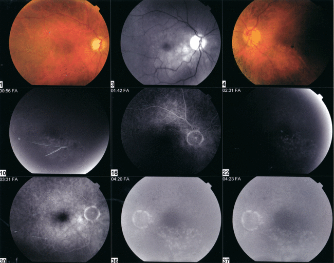
Fluorescein angiography of the retina in each eye
Discussion:
The lack of perfusion abnormalities of the right eye is consistent with a possible non-ischemic central retinal vein occlusion in the right eye. The pallor of the optic disc and attenuation of blood vessels suggest likely glaucomatous optic atrophy associated with the vein occlusion in the right eye. There is no evidence of macular edema or neovascularization of the right eye, and the only finding of significance was a mild epiretinal membrane in the right eye that can be observed on examination.
In the left eye, there is evidence of prior branch retinal vein occlusion treated with focal laser. The superior hemiretina of the left eye is of concern and most likely represents moderate branch vein occlusion in evolution. However, it is not visually significant at the time of fluorescein angiography and is manifested only as a moderate intraretinal hemorrhage, There is no associated macular edema in the left eye.
Conclusion:
This patient had evidence of prior ocular problems that was discovered on forensic analysis of her medical records and documented through fluorescein angiography of the retina. Laser treatment of the retina produces defects of the retinal pigment epithelium that may be diagnosed on fluorescein angiography. Forensic analysis by fluorescein angiography also showed a pale optic nerve that was consistent with glaucoma associated with a central retinal vein occlusion. Forensic analysis was able to show ophthalmic conditions that were inconsistent with her history of vision loss.
Return to Home Page from Forensic Analysis of Vision Loss Following a Motor Vehicle Accident